
UCSF Identifies New Cause of Neurologic Disease, Saves a Teenager's Life
Five months after receiving a kidney transplant, a teenage girl arrived at UCSF Benioff Children's Hospital San Francisco with headaches, low sodium levels, and worsening kidney function. Imaging showed concerning inflammation involving structures deep in the brain and behind her eyes.
Her care team faced an urgent question: was her immune system attacking her own tissue, or was an unidentified infection attacking it instead? The distinction mattered enormously, because suppressing her immune system could calm an autoimmune flare, but could also let an infection spread unchecked.
An extensive workup, including a biopsy of the pituitary gland itself, found no clear answer, and her condition kept worsening.
Erica Winnicki, MD, the pediatric nephrologist overseeing her transplant care, continued to search for an answer. "Every standard test we ran failed to identify an infection, but her condition kept telling us we were missing something," Winnicki said.

A Virus Nobody Expected
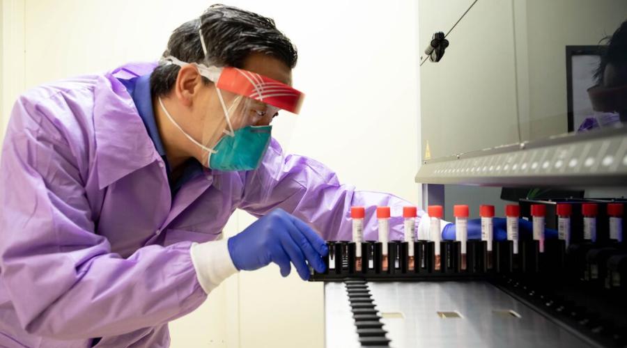
The team turned to metagenomic sequencing, an advanced test that reads all the genetic material in a sample rather than checking for one suspected germ at a time. The analysis, run by Charles Chiu, MD, PhD, of UCSF's Clinical Microbiology Laboratory, turned up a virus called trichodysplasia spinulosa–associated polyomavirus (TSPyV). This virus was unexpected: it was known only for causing a skin condition.
"We found a virus, but that wasn't enough," said Chiu. "This virus had never been known to be invasive, so we needed to prove that what we were seeing was real, and that it explained what was happening to our patient."
Confirming the Culprit
As the investigation continued, her condition grew more serious. Vision loss, hearing loss, and worsening brain function required additional time in intensive care.
Further laboratory testing found evidence of TSPyV in tissue from her pituitary gland and kidney, while analysis of her immune response pointed toward an active viral infection rather than noninfectious inflammation. Electron microscopy from the laboratory of Joseph DeRisi, PhD, a co-author on the paper, revealed virus particles consistent with a polyomavirus like TSPyV.
"Once we could see the virus itself at the base of her brain, and her body's own immune response confirmed it was fighting a viral infection, we had our answer for this brain disease," said Kendall Nash, MD, a pediatric neurologist on the care team.
Recovery — and a New Diagnostic Lead
With the diagnosis confirmed, the team reduced the patient’s immunosuppressive medications and started antiviral treatment. Within two weeks, her vision returned to 20/20, her mental status returned to baseline, and her headaches resolved. In total, she was well enough to go home after nearly 80 days in the hospital.
Several months later, her kidney function had improved enough to come off dialysis therapy and she had resumed life as a busy high school student.
The case — first authored by Aaron Bodansky, MD, a pediatric critical care physician on the care team — was described in the New England Journal of Medicine and is the first to implicate TSPyV as a cause of invasive neurologic disease. For physicians treating immunocompromised patients with unexplained neurologic decline, it adds a new possibility to consider when standard testing comes up empty.
“We saved a teenager's life by discovering a neurologic virus in real time,” Bodansky said. “We took that discovery straight to her bedside — and now other physicians know to look for this virus, too. This is what makes UCSF such a special place.”
Authors: Other authors from the UCSF Department of Pediatrics are Thomas Tarro, MD, Nahid Hiermandi, MD, William Bernal, MD, MPH, Alexandra Bicki, MD, MPH, Marsha Lee, MD, and Matt Zinter, MD. For all authors, see the paper.
Funding: This work was supported by grant 1U01AT012993 from the National Institutes of Health and by the Chan Zuckerberg Biohub.